


Building Better Clinical Evidence with Medical Imaging Data
Introduction
Clinical trials depend on evidence that is accurate, measurable, and consistent. Every study outcome depends on the quality of the data collected from participants. While lab values, clinical assessments, safety reports, and patient-reported outcomes are important, imaging provides a powerful visual layer of evidence. This is why medical imaging in clinical trials has become a critical part of modern research across oncology, neurology, cardiology, orthopedics, respiratory diseases, and other therapeutic areas.
Imaging allows study teams to observe disease progression, treatment response, anatomical changes, and functional improvements. It helps sponsors, CROs, investigators, radiologists, and imaging core labs make more informed decisions during the trial. As studies become more data-driven, clinical trial imaging is playing a stronger role in patient selection, endpoint evaluation, and treatment monitoring.
Why Medical Imaging Is Valuable in Clinical Trials
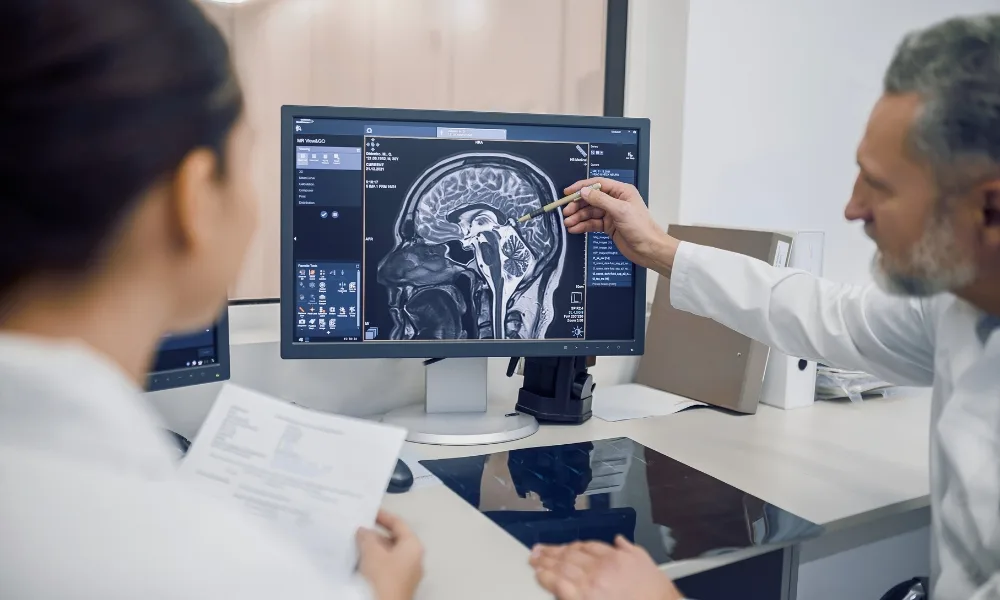
Medical imaging in clinical trials helps research teams collect objective information about a participant’s condition. Imaging techniques such as CT, MRI, PET, ultrasound, and X-ray can show tumors, lesions, organ changes, tissue damage, blood flow, inflammation, and structural abnormalities.
In oncology studies, imaging is often used to measure tumor burden and assess whether a therapy is working. In neurology trials, MRI can help monitor lesions, brain structure, or disease progression. In cardiology research, imaging may be used to evaluate heart function, vascular health, or blood flow. In orthopedic trials, imaging can help assess bone healing, joint damage, or tissue repair.
Because imaging provides measurable visual evidence, it can strengthen clinical trial conclusions and support more reliable decision-making.
Clinical Trial Imaging for Patient Selection
Selecting the right participants is essential for any successful clinical trial. If patients do not meet the protocol criteria, the quality of the study data may be affected. Clinical trial imaging can help confirm eligibility before enrollment.
For example, an oncology trial may require measurable disease at baseline. A neurology study may need MRI evidence of a specific disease stage. A cardiovascular study may require imaging confirmation of heart or vessel conditions.
By using imaging during screening, trial teams can ensure that participants match the study requirements. This improves enrollment accuracy and helps reduce protocol deviations.
Imaging for Treatment Response Assessment
One of the most important uses of medical imaging in clinical trials is treatment response assessment. Imaging allows researchers to compare baseline scans with follow-up scans to see whether a disease is improving, stable, or progressing.
In oncology, standardized criteria such as RECIST may be used to measure tumor response. Radiologists compare lesions over time and classify the response based on defined rules. In other therapeutic areas, imaging may help measure inflammation, tissue repair, organ function, or structural changes.
This makes imaging especially useful when symptoms or lab results alone do not provide a complete picture of treatment effect.
Why DICOM Is Important in Clinical Trial Imaging
As imaging became more central to clinical trials, the need for standardization increased. This is where DICOM in clinical trials becomes important. DICOM stands for Digital Imaging and Communications in Medicine. It is the standard format used to store, exchange, and manage medical images.
DICOM medical imaging includes both the image and important metadata. This metadata may include scan date, imaging modality, scanner details, image orientation, acquisition parameters, patient identifiers, and study-related information.
In clinical trials, imaging data may come from multiple sites, scanners, hospitals, and countries. Without a common standard, it would be difficult to organize, transfer, review, and analyze imaging data consistently. DICOM helps create a structured workflow for managing imaging data across the study.
DICOM Medical Imaging and Trial Traceability
Traceability is essential in clinical research. Every image should be connected to the correct participant, visit, timepoint, and study. DICOM medical imaging supports this by preserving important technical and study-related information within the image file.
For example, if a protocol requires a specific MRI sequence or CT acquisition setting, DICOM metadata can help reviewers verify whether the scan meets the required standard. If metadata is missing or incorrect, the image may require follow-up before it can be reviewed.
Proper use of DICOM in clinical trials helps sponsors and CROs improve image tracking, reduce review delays, and maintain stronger data quality.
Common Challenges in Imaging Data Management
Although clinical trial imaging provides important evidence, it can be difficult to manage. Imaging files are large and require secure upload, storage, transfer, and review. Studies may also require repeated scans across multiple visits, increasing the volume of data.
Another challenge is site variation. Different trial sites may use different scanners, acquisition settings, or workflows. If imaging protocols are not followed consistently, image comparability may be affected.
De-identification is also critical. DICOM files may contain patient information in metadata fields. Before images are shared for central review or analysis, patient identifiers must be removed or masked while preserving essential study details.
Strong imaging workflows help manage these challenges by standardizing image capture, quality control, anonymization, transfer, and review.
The Role of Central Imaging Review
Many clinical trials use central imaging review to improve consistency and reduce bias. In this process, images from different sites are reviewed by independent radiologists or imaging specialists using predefined criteria.
Central review is especially important when imaging contributes to primary or secondary endpoints. It helps ensure that images are interpreted consistently across the trial, regardless of where they were captured.
A reliable central review process depends on complete, high-quality, properly de-identified imaging data. Strong DICOM medical imaging workflows help ensure that images are review-ready and traceable.
How AI Is Supporting Medical Imaging Workflows
AI is increasingly being used to support imaging workflows in clinical trials. It can help with image quality checks, lesion detection, segmentation, measurement support, anonymization review, and imaging biomarker analysis.
AI can also help manage large imaging datasets by identifying patterns or flagging scans that may need closer review. However, AI depends on standardized and high-quality imaging data. This makes DICOM in clinical trials and strong imaging governance even more important.
AI should support radiologists and imaging experts, not replace them. Human expertise remains essential for final interpretation, validation, and clinical decision-making.
Conclusion
Medical imaging in clinical trials helps research teams build stronger clinical evidence by supporting patient selection, disease monitoring, treatment response assessment, and endpoint evaluation. It provides objective visual data that can improve confidence in study outcomes.
DICOM in clinical trials provides the standard structure needed to manage imaging data across sites, scanners, systems, and reviewers. With strong DICOM medical imaging workflows, sponsors and CROs can improve traceability, image quality, and review consistency.
As clinical research becomes more complex, strong clinical trial imaging processes will be essential for turning imaging data into reliable clinical evidence.
Categorias
Leia Mais
テクノロジーが飛躍的な進化を遂げ、あらゆるエンターテインメントが手のひらの中で完結する2026年、私たちの娯楽の形もかつてないほど豊かになりました。多くの愛好家が注目しているオンラインカジノ 合法的で安全なサービスを追求する動きは、安心してゲームを楽しむための重要なスタンダードとなっています。PCやスマートフォンからいつでもアクセス可能な環境は、忙しい日常に彩りとワクワク感をもたらしてくれます。本記事では、オンラインカジノが持つ独自の魅力や、初めての方でも安心して楽しむためのポイントを詳しくご紹介します。 ライフスタイルに調和するオンラインカジノの利便性...

The stage is set for a historic Pacific showdown in Santiago. ' For the first time ever, Nongshim RedForce and Paper Rex will clash, this time with everything on the line. ' While a prediction is inherently speculative, Paper Rex may hold a slight edge due to their deep reservoir of grand finals experience. ' The PRX squad, featuring the dynamic flexibility of f0rsaken and jinggg alongside the...

Muscle pain and stiffness are common experiences that can affect people of all ages. These issues may arise from physical activity, prolonged sitting, minor injuries, or even daily stress. When muscles feel tight or sore, it can limit movement and make simple activities uncomfortable. Over time, ignoring these signs may lead to increased discomfort and reduced mobility. Because of this, finding...

NEWARK, DELAWARE | April 6, 2026 — The demand for dehydrated onions in the European Union is witnessing rapid expansion, driven by strong growth in food processing activities and increasing reliance on shelf-stable ingredients. Valued at USD 470.70 million in 2025, the market is projected to reach USD 545.54 million in 2026 and is forecast to surge to USD 2.39 billion by...

Learning a new language can open doors to better travel experiences, career opportunities, cultural understanding, and personal growth. However, many learners struggle to find a platform that goes beyond simple vocabulary memorization and actually teaches real communication skills. That’s where Rocket Languages stands out. Rocket Languages is one of the most recognized online language...